
click on the images to jump to some results
While most neurons are generated during the embryonic and postnatal periods, it is now well accepted that some regions of the brain keep producing new neurons throughout adulthood. Understanding the processes that govern the birth and development of neurons could lead to future treatments to a variety of pathological and neurodegenerative conditions using adult neural stem cells. In order to understand these processes, neuroscientists need to study the movement and morphology of neurons as they migrate and develop. We have developed state-of-the-art methods for detecting the nuclei of developing neurons and tracking them as they migrate within the brain.
K. Smith, K. Ali, A. Carleton, V. Lepetit
Results
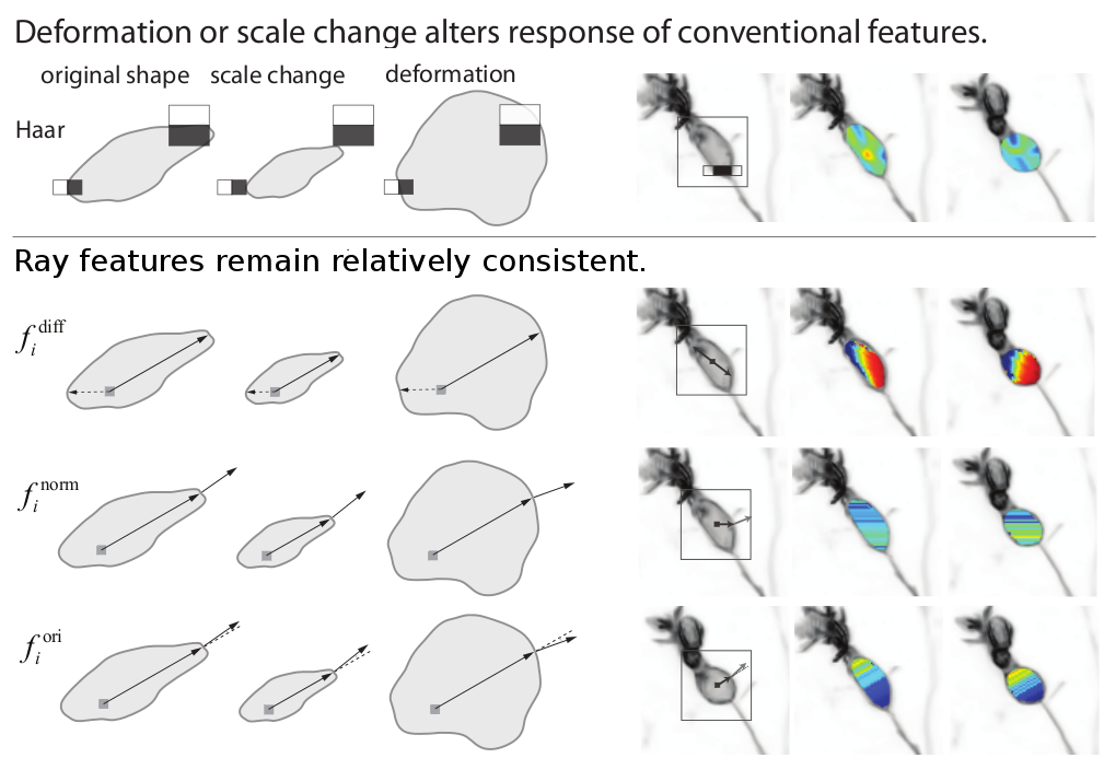
To detect neurons in microscopy data, we developed a detector which learns the appearance of neurons, even though it may vary greatly from neuron to neuron. An Adaboost learning framework is used with a new class of image features called Rays. Rays are capable of characterizing deformable or irregular shapes such as neurons with consistent responses, whereas standard features including Haar and HOG are less reliable. They can also provide contextual information outside of the detector window.
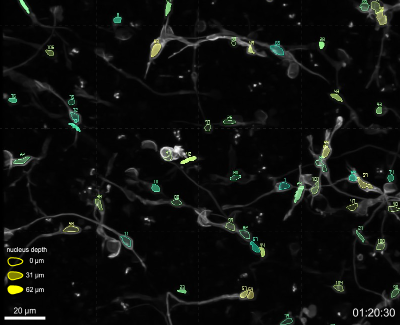
Our neuron detector is applied to 2-photon time-lapse microscopy data, containing hundreds of migrating neurons. Each neuron is tracked by linking the detections using a batch MCMC data association approach that estimates the MAP of a Bayesian filtering distribution. Two general constraints help us limit the search space. The first constraint relaxes the standard assumption that target motion and appearance are independent, and learns a correlation between the appearance of a neuron and its motion – the neuron nucleus elongates in the direction of motion (increasing elongation with speed). The second constraint forces neurons to enter and exit the scene at image boundaries.
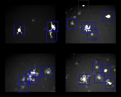
We have also developed methods to track neurons and reduce the amount of labeling necessary. In the video on the right, migrating neurons are tracked and automatically annotated. Users have clicked on the neuron locations in 1/32 of the frames, denoted by cyan bounding boxes. In all other frames, the neuron is detected and tracked automatically. Results are shown by the blue bounding boxes. Gray bounding boxes indicate the true location of the neurons.
Materials
References
Please note that the publication lists from Infoscience integrated into the EPFL website, lab or people pages are frozen following the launch of the new version of platform. The owners of these pages are invited to recreate their publication list from Infoscience. For any assistance, please consult the Infoscience help or contact support.
Fast Ray Features for Learning Irregular Shapes
2009. 12th IEEE International Conference on Computer Vision, Kyoto, JAPAN, Sep 29-Oct 02, 2009. p. 397-404. DOI : 10.1109/ICCV.2009.5459210.Please note that the publication lists from Infoscience integrated into the EPFL website, lab or people pages are frozen following the launch of the new version of platform. The owners of these pages are invited to recreate their publication list from Infoscience. For any assistance, please consult the Infoscience help or contact support.
General Constraints for Batch Multiple-Target Tracking Applied to Large-Scale Videomicroscopy
2008. IEEE Conference on Computer Vision and Pattern Recognition, Anchorage, AK, June 2008. DOI : 10.1109/CVPR.2008.4587506.Contacts
| Kevin Smith | [e-mail] |
| Karim Ali | [e-mail] |
| Alan Carleton | [e-mail] |
| Vincent Lepetit | [e-mail] |