Pancreatic ductal adenocarcinoma is a devastating type of cancer with poor prognosis and low survival rates. In it, the typically soft (stroma) surrounding the tumor site stiffens into a dense, fibrous tumor microenvironment[1]. This stiffening, which seems to block the uptake and effectiveness of anticancer drugs, is caused by the crosstalk between pancreatic cancer cells and the surrounding stromal cells. To better understand this inflammatory process, we aim to bioprint 3D models of pancreatic tissue and follow the molecular interaction across cells within them.
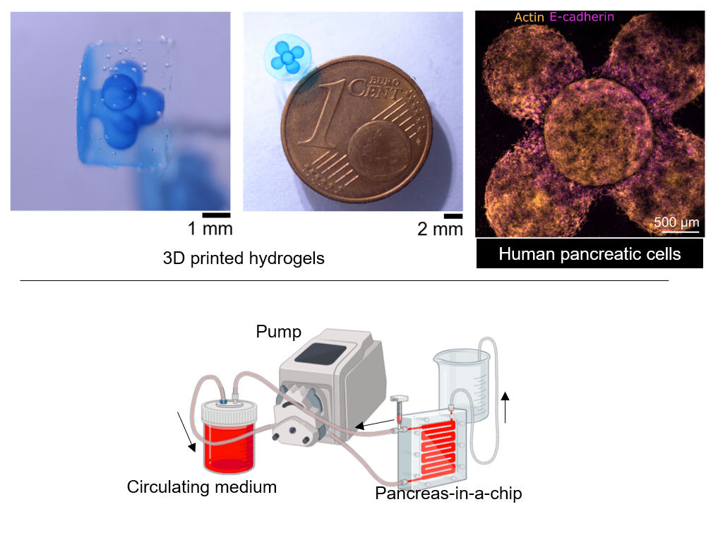
We use a novel light-based bioprinting technology developed in our lab to fabricate these 3D tissue models. This technology, called tomographic volumetric bioprinting[2], allows us to fabricate cell-laden cm-scale complex structures within seconds. These bioprinted constructs are highly viable and can be matured for up to several weeks[3].
The objective of this master’s project is to contribute to the development of more biologically relevant biofabricated pancreatic adenocarcinoma models by incorporating vasculature for nutrients diffusion. These complex constructs, populated by multiple cell types, can then be used for drug screening and interrogated with advanced microscopy techniques to study cell behavior.
Project Goals:
1. Design and Fabricate a Perfusion System: The primary task will be to design and fabricate a perfusion system to pump nutrients through the bioprinted tissue models. This system should be leak-free, robust, and sterilizable to ensure consistent and reliable nutrient delivery to the cells. Ideally, we should be able to perfuse multiple tissue models in parallel.
2. Manufacture micrometric channels for vasculature by 3D printing: Incorporate vasculature[4] into the bioprinted pancreatic cancer models to create more biologically relevant constructs.
Contact
Viola Sgarminato ([email protected])
Jorge Madrid Wolff ([email protected])
References
1. Orth, M. et al. Pancreatic ductal adenocarcinoma: Biological hallmarks, current status, and future perspectives of combined modality treatment approaches. Radiation Oncology vol. 14 1–20 Preprint at https://doi.org/10.1186/s13014-019-1345-6 (2019).
2. Madrid-Wolff, J. et al. A review of materials used in tomographic volumetric additive manufacturing. MRS Commun 13, 764–785 (2023).
3. Sgarminato, V. et al. 3D in vitro modeling of the exocrine pancreatic unit using tomographic volumetric bioprinting. bioRxiv (2024) doi:10.1101/2023.01.23.525142.
4. Mair, Vincent, et al. “Freeform printing of thermoresponsive poly (2-cyclopropyl-oxazoline) as cytocompatible and on-demand dissolving template of hollow channel networks in cell-laden hydrogels.” Biofabrication 14.2 (2022): 025019.