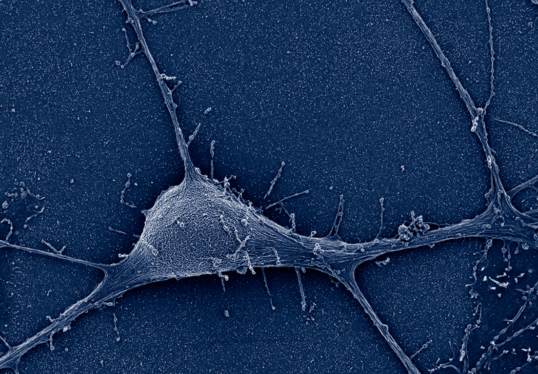
 Image of a neuron using a SEM
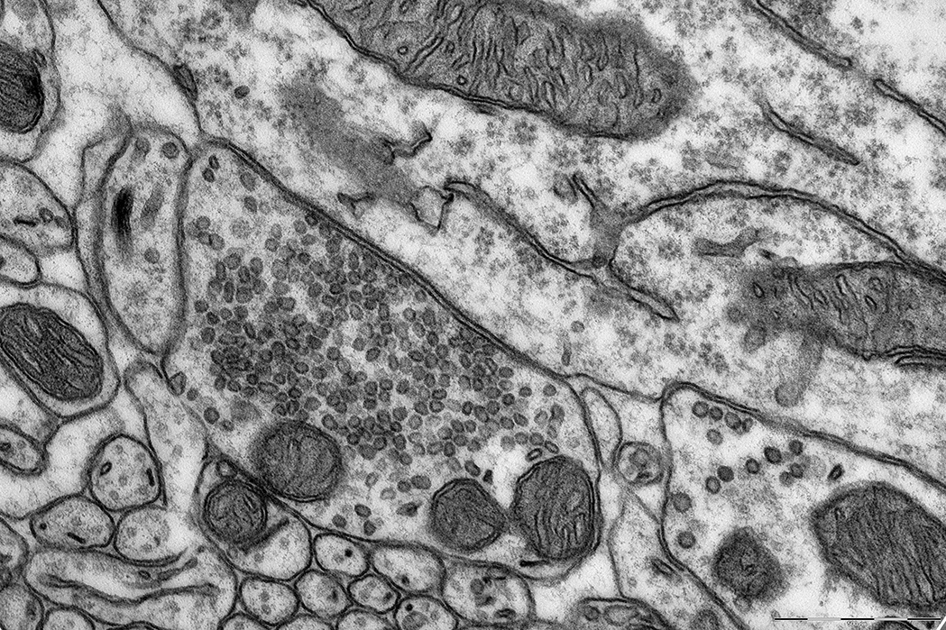 TEM image of an axon
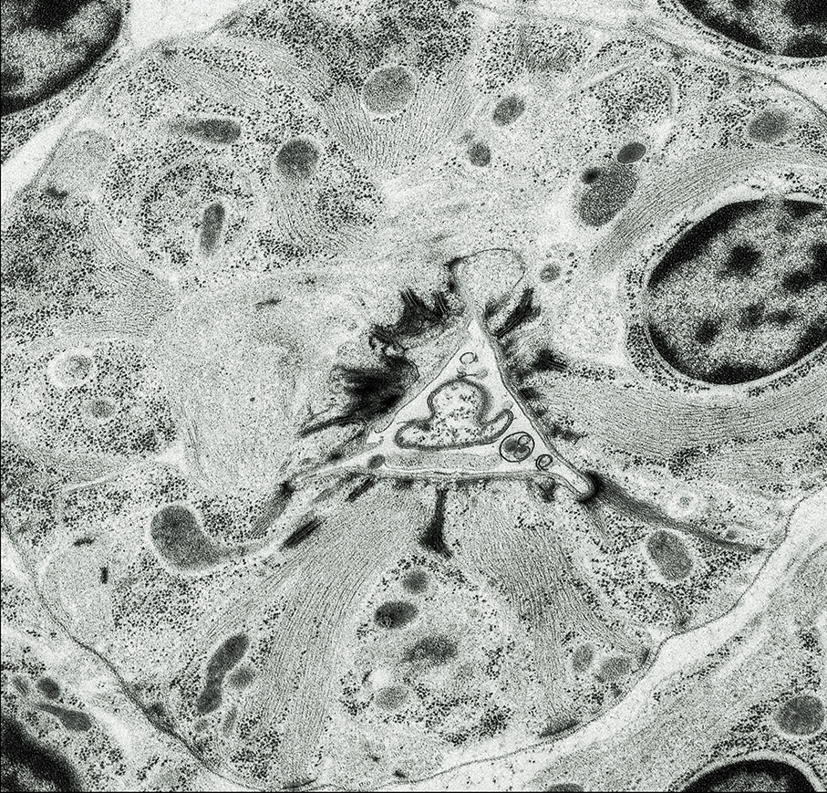
TEM image of a worm
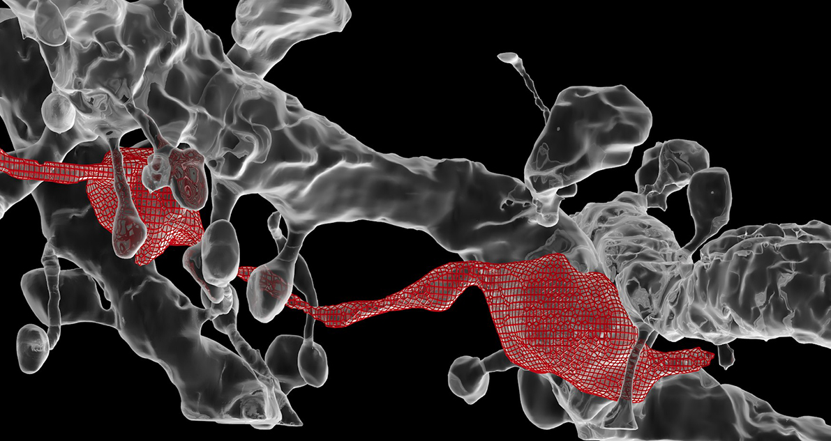 3D reconstruction of an axon (red) and a dentrite (grey) using Blender
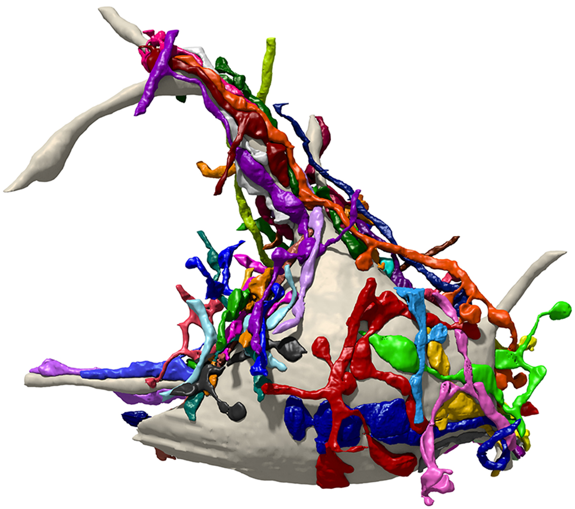
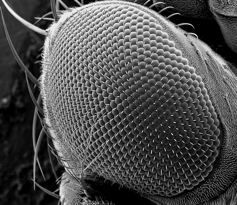 SEM of a fly eye
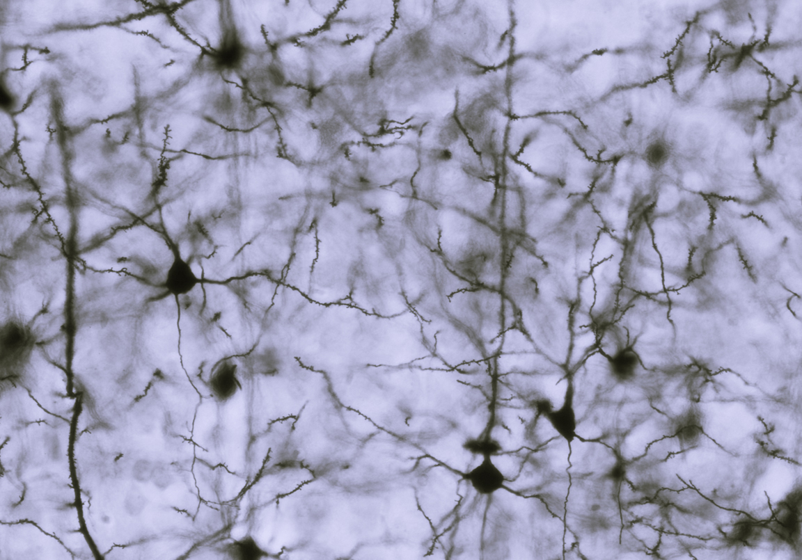 Neurons
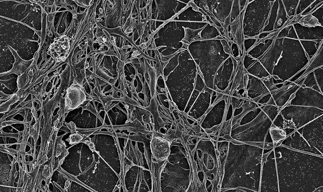 SEM of neurons
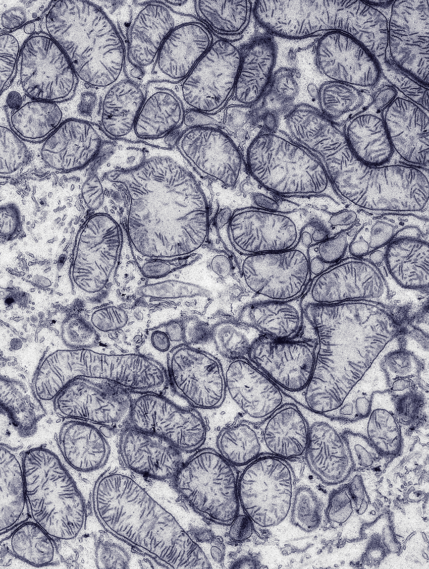 TEM image of mitochondria
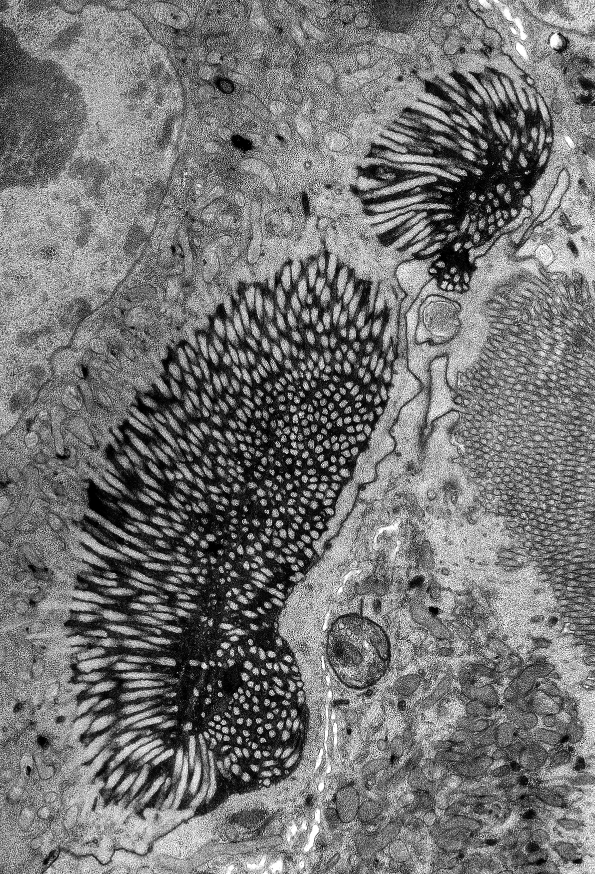
Rough ER of a worm
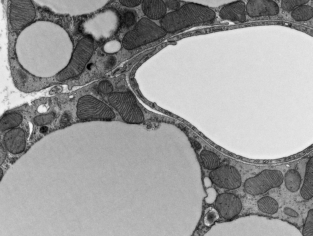
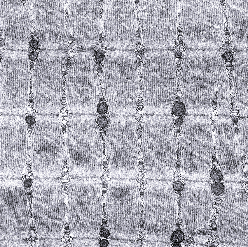
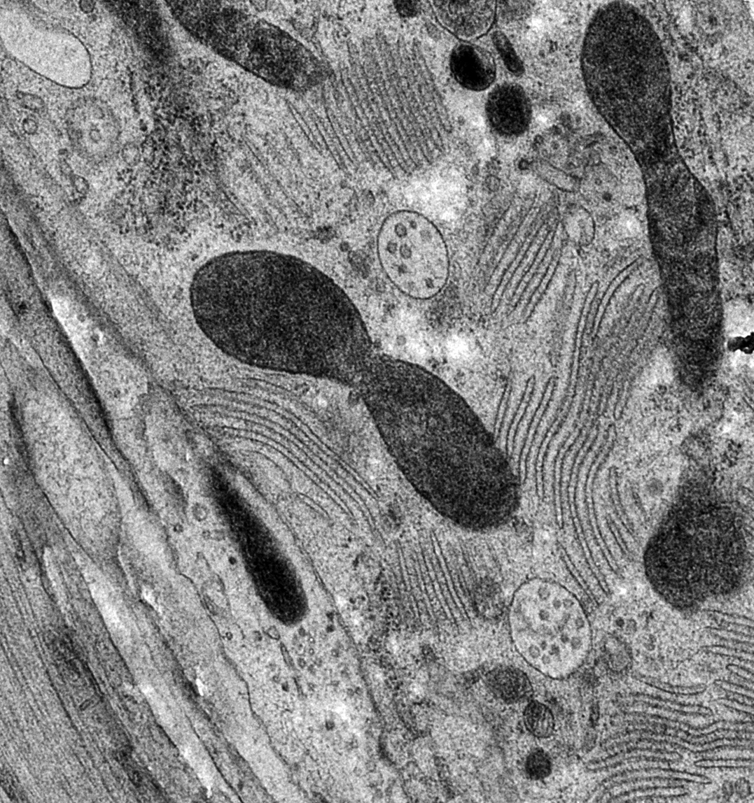
TEM image of muscle tissue
 3D reconstruction of an axon using the NeuroMorph plugin in Blender. EM images were obtained using a Serial Block-Face Scanning Electron Microscope (SBEM).
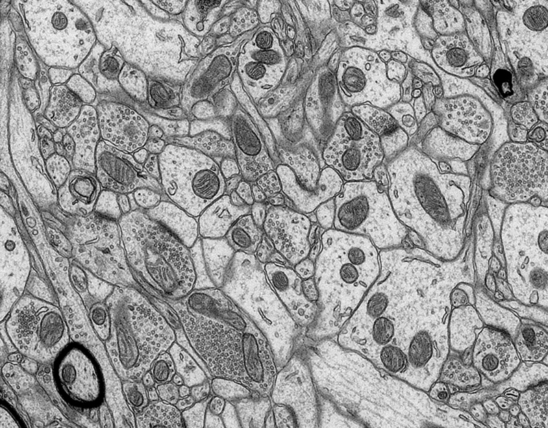
TEM image of brain tissue
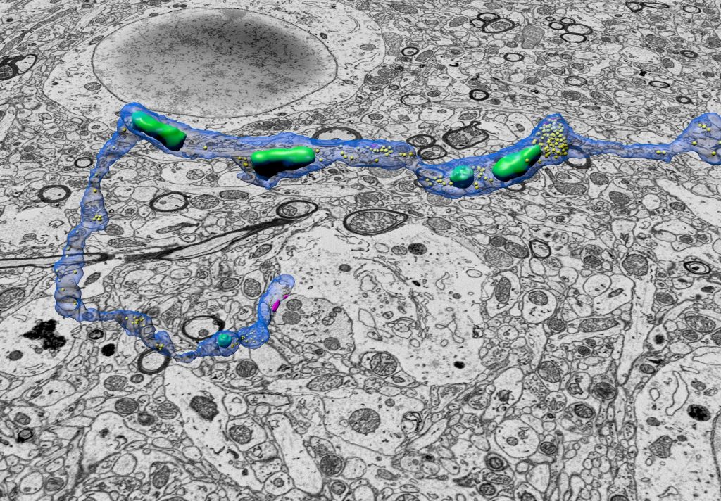
3D reconstruction of neurons using the NeuroMorph plugin in Blender. EM images were obtained using a Focused Ion Beam Scanning Electron Microscope (FIB-SEM).
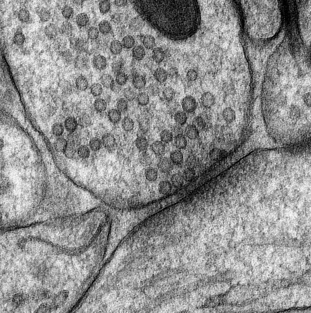
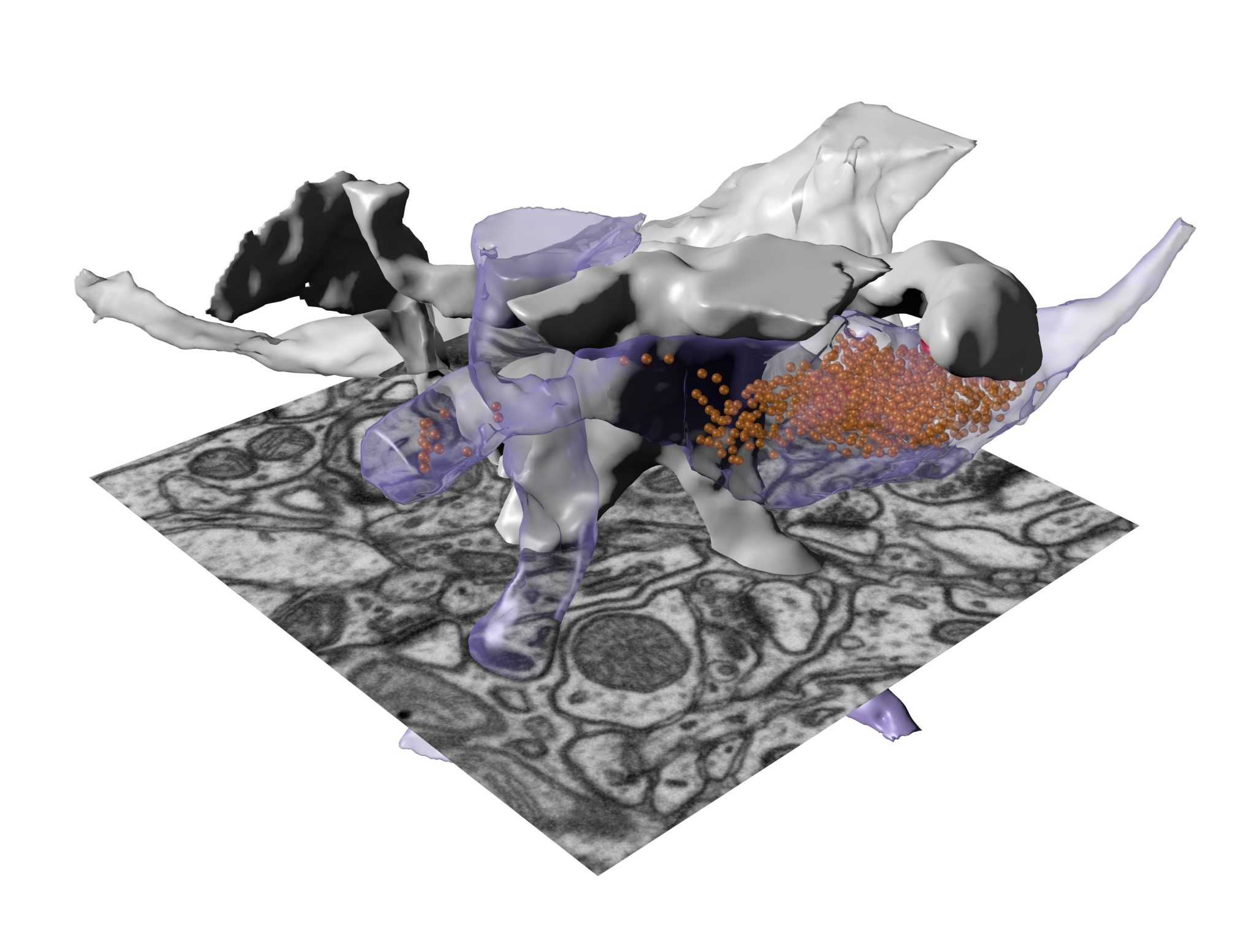
TEM image of a synapse