November/December 2022 competitors
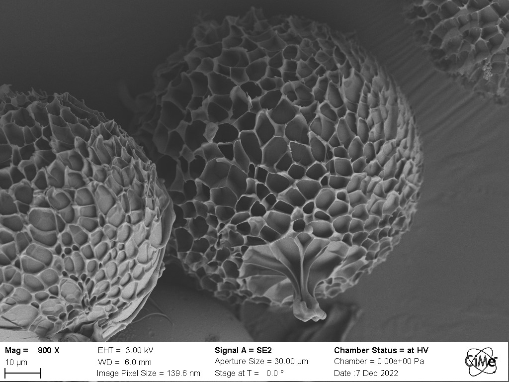
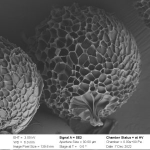
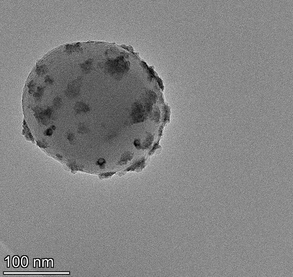
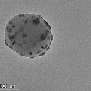
Freeze-dried polymeric core shell particle. Microscope: SEM Gemini. 1/8
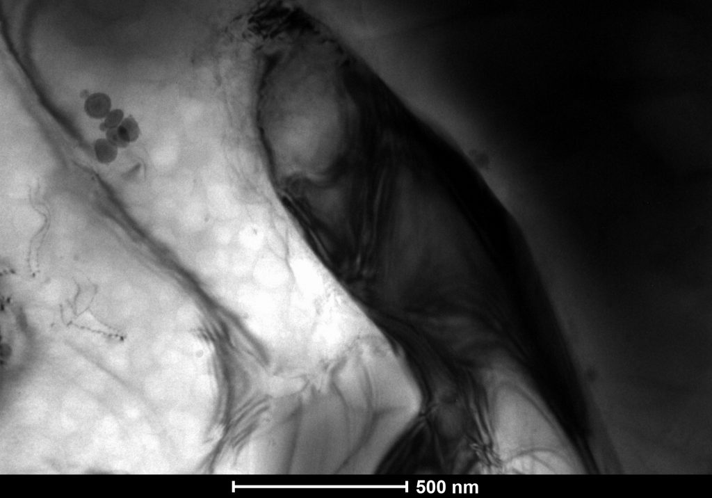
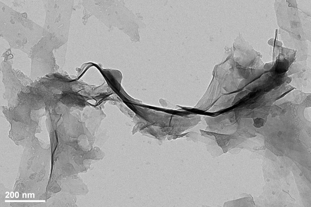
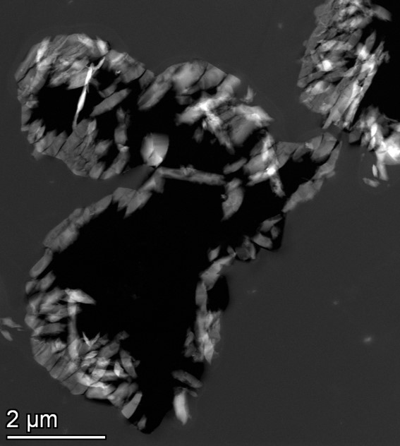
Bright-field TEM image of Ni3Al alloy. Microscope: TEM Osiris. 2/8
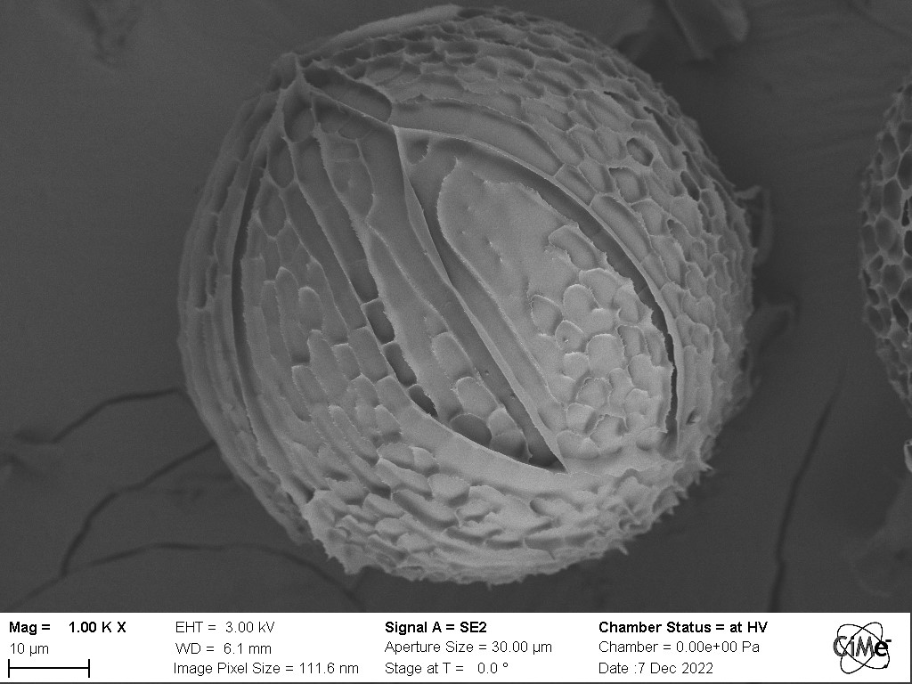
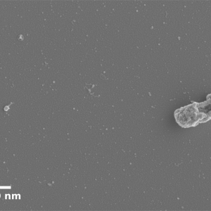
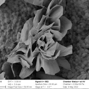
Freeze-dried core shell particles composed of polymers. Microscope: SEM Gemini. 3/8
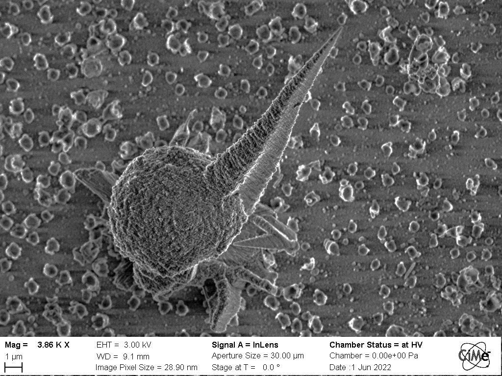
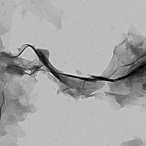
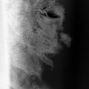
After a long hard fight, Alicia, the bacterium, did not survive Alex’s attack, the bacteriophage. After spending some minutes in salty water, Alex, with his long tail (~150 nm), bound to Alicia’s cell wall. His spike perforated the membrane, allowing him to inject his DNA into its host: virion replication started. The bacterium realized after some time that it was game over. Her membrane started to tear up when a sudden burst released tens of bacteriophages in the environment. The result is ruthless. Special thanks to Mary-Claude Croisier for the sample preparation. Microscope: SEM Merlin. 4/8
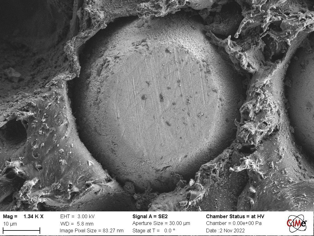
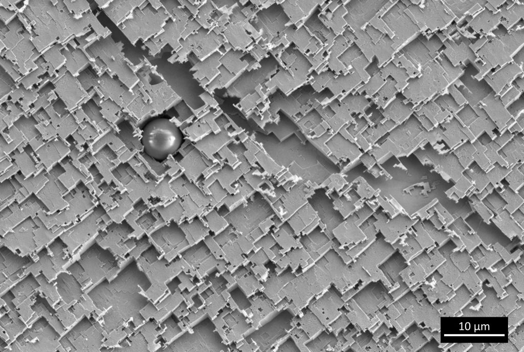
In addition to the visible scratches on the metallic particles and the surface fraying of the surrounding thermoplastic, the polishing process resulted in enlarging of the cavity around the particle, wrongly suggesting a bad adhesion between both materials. Microscope: SEM Gemini. 5/8
Freeze-dried polymeric core shell particle. Microscope: SEM Gemini. 6/8
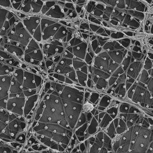
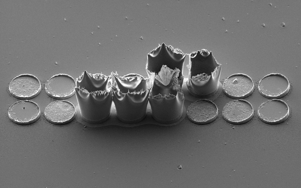
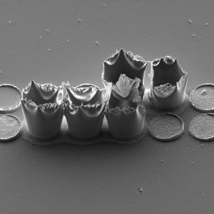
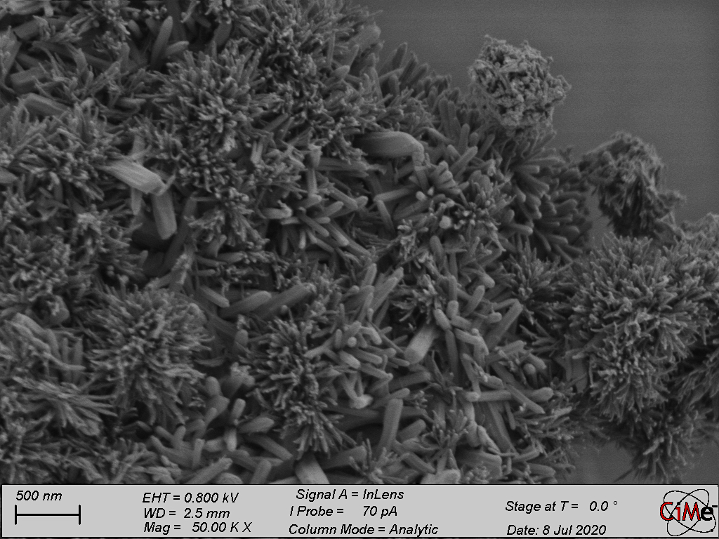
Micro Copper structure electrodeposited by controlling the current density. Microscope: SEM Merlin. 7/8
Micro Copper structure electrodeposited by controlling the current density. Microscope: SEM Merlin. 8/8
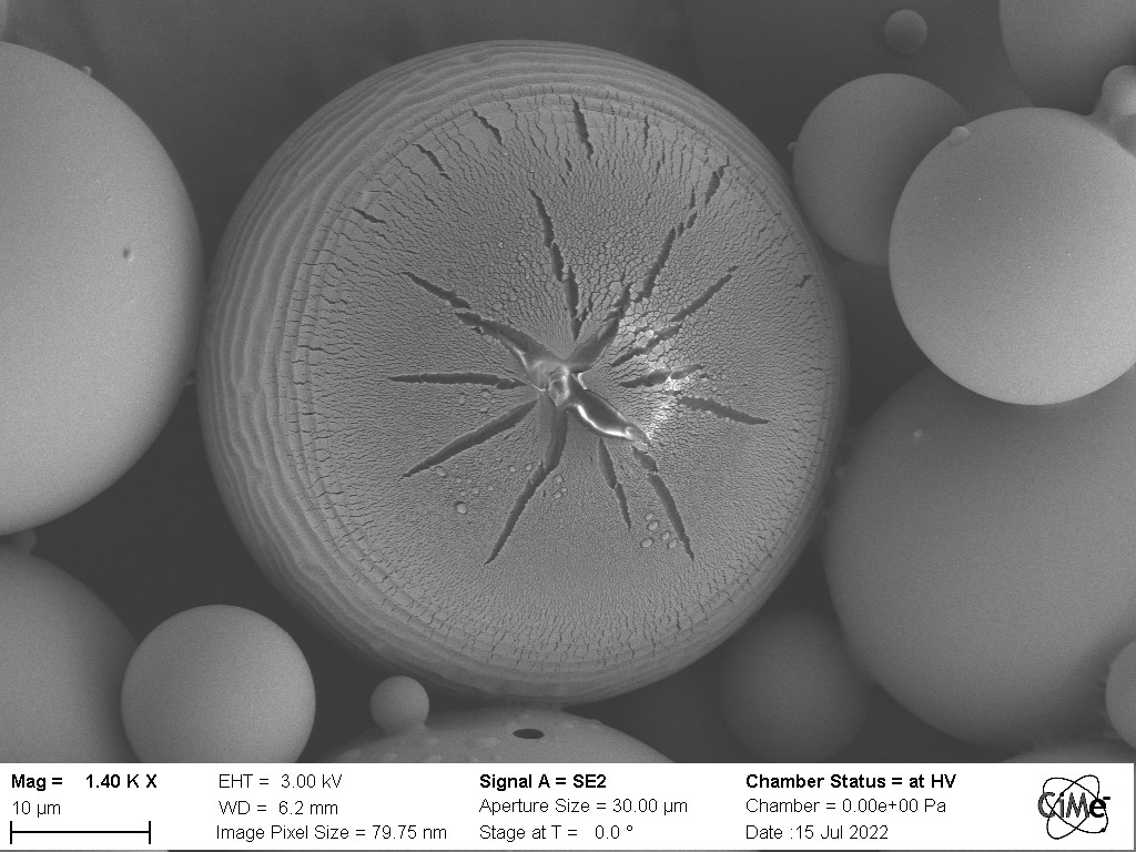
Freeze-dried polymeric core shell particle. Microscope: SEM Gemini.
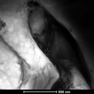
Bright-field TEM image of Ni3Al alloy. Microscope: TEM Osiris.
Freeze-dried core shell particles composed of polymers. Microscope: SEM Gemini.
After a long hard fight, Alicia, the bacterium, did not survive Alex’s attack, the bacteriophage. After spending some minutes in salty water, Alex, with his long tail (~150 nm), bound to Alicia’s cell wall. His spike perforated the membrane, allowing him to inject his DNA into its host: virion replication started. The bacterium realized after some time that it was game over. Her membrane started to tear up when a sudden burst released tens of bacteriophages in the environment. The result is ruthless. Special thanks to Mary-Claude Croisier for the sample preparation. Microscope: SEM Merlin.
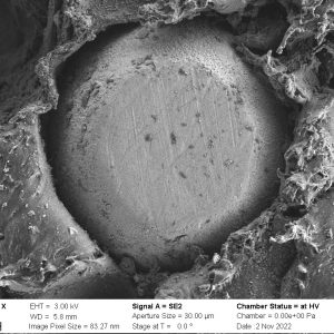
In addition to the visible scratches on the metallic particles and the surface fraying of the surrounding thermoplastic, the polishing process resulted in enlarging of the cavity around the particle, wrongly suggesting a bad adhesion between both materials. Microscope: SEM Gemini.
Freeze-dried polymeric core shell particle. Microscope: SEM Gemini.
Micro Copper structure electrodeposited by controlling the current density. Microscope: SEM Merlin.
Micro Copper structure electrodeposited by controlling the current density. Microscope: SEM Merlin.
September/October 2022 competitors
Micro Copper structure electrodeposited by controlling the current density. Microscope: SEM Merlin. 1/7
The crystal from etching solution stays on the graphene lattice when preparing the graphene specimen. The crystal is surrounded by contamination. Microscope: TEM Themis.
2/7
Micro Copper structure electrodeposited by controlling the current density. Microscope: SEM Merlin. 3/7
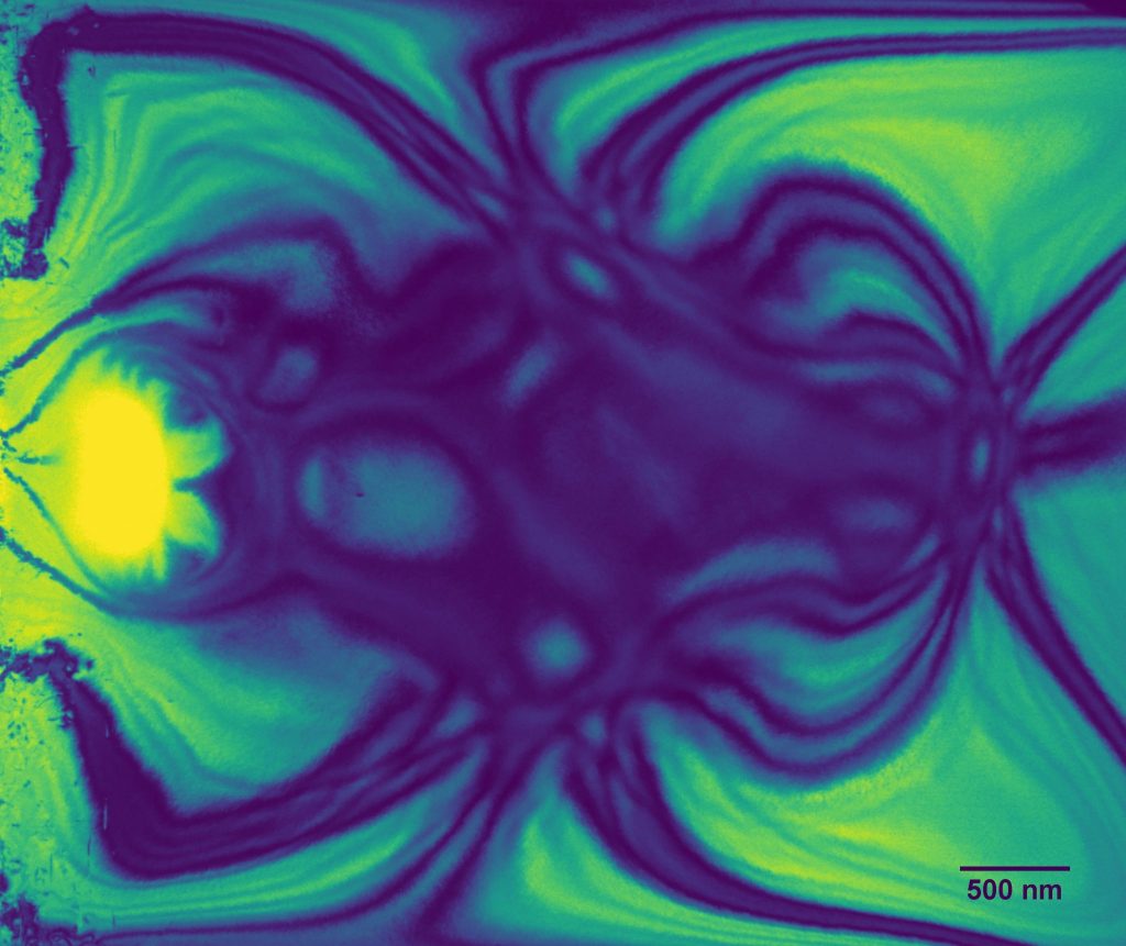
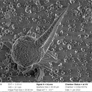
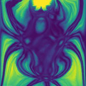
Potassium niobate lamella prepared using focused ion beam. As an effect of the bend contours a spider appears on the surface. Microscope: TEM Talos. 4/7
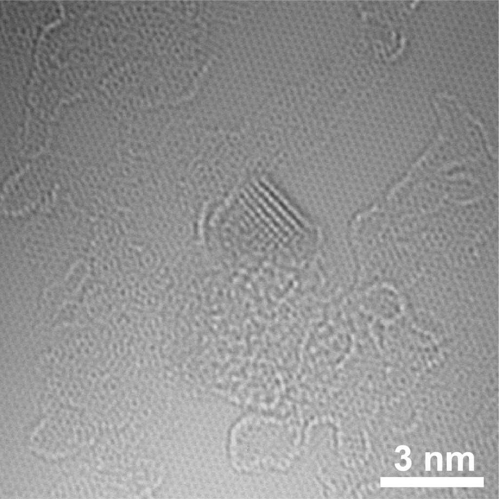
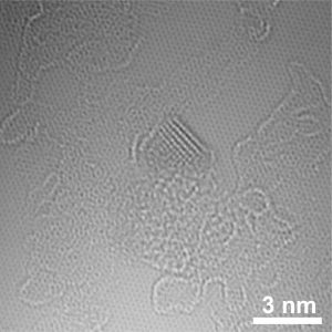
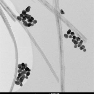
This semiconducting nanoparticle is decorated with metal oxide and pure metal catalysts. Microscope: TEM Talos. 5/7
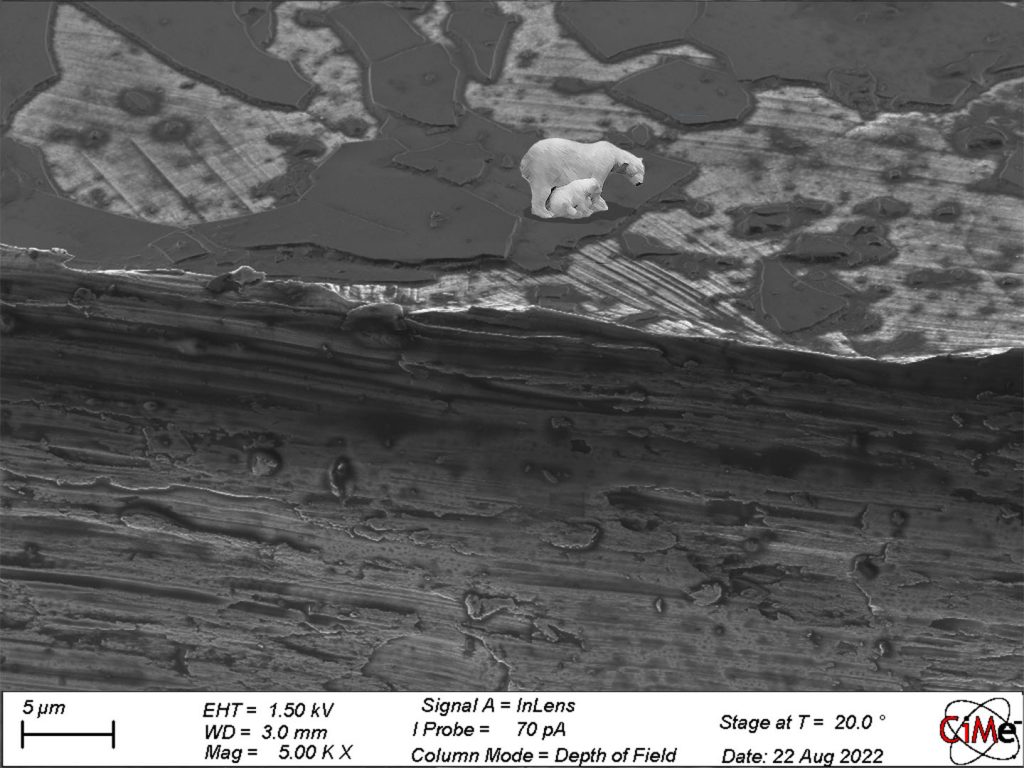
Image of the broken carbon membranes on nickel makes me think of the severe global warming effect and the melting planet for poor polar bears. Microscope: SEM Merlin. 6/7
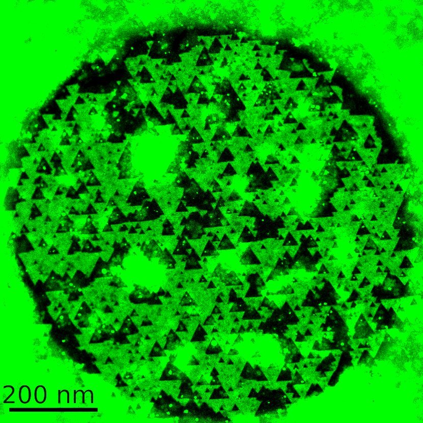
Triangular pores arranged in WS2 like Christmas trees. Microscope: TEM Themis. 7/7
Micro Copper structure electrodeposited by controlling the current density. Microscope: SEM Merlin.
The crystal from etching solution stays on the graphene lattice when preparing the graphene specimen. The crystal is surrounded by contamination. Microscope: TEM Themis.
Micro Copper structure electrodeposited by controlling the current density. Microscope: SEM Merlin.
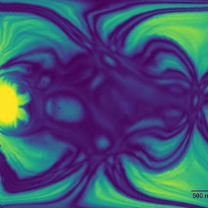
Potassium niobate lamella prepared using focused ion beam. As an effect of the bend contours a spider appears on the surface. Microscope: TEM Talos.
This semiconducting nanoparticle is decorated with metal oxide and pure metal catalysts. Microscope: TEM Talos.
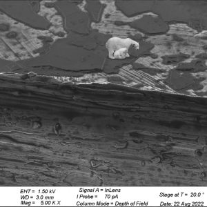
Image of the broken carbon membranes on nickel makes me think of the severe global warming effect and the melting planet for poor polar bears. Microscope: SEM Merlin.
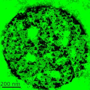
Triangular pores arranged in WS2 like Christmas trees. Microscope: TEM Themis.
July/August 2022 competitors
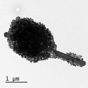
Polymeric microparticles processed via vortexing. Microscope: SEM Gemini. 1/4

Nothing better than a nano ice cream during a hot summer afternoon. Composed of randomly distributed Cu nanocrystals around a Cu rod. Microscope: TEM Tecnai G2 Spirit Twin (Sion). 2/4
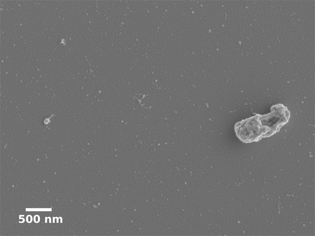
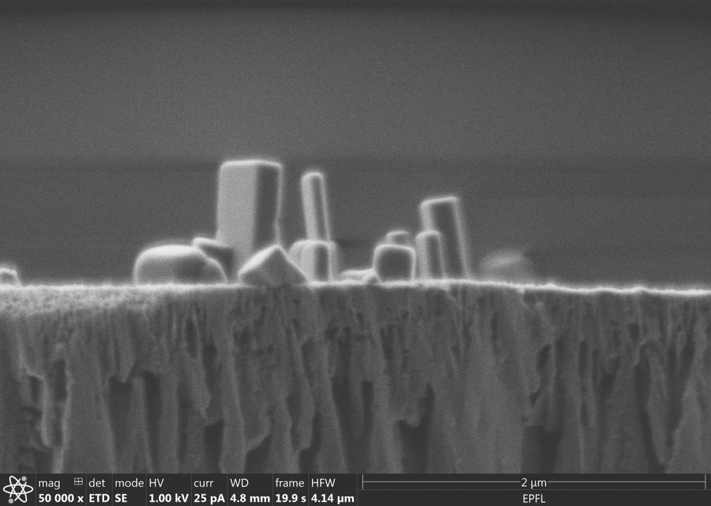
The probe apex (made by Pt/Ir wire) for the scanning tunneling microscope (Sion). Microscope: FEI Teneo SEM. 3/4
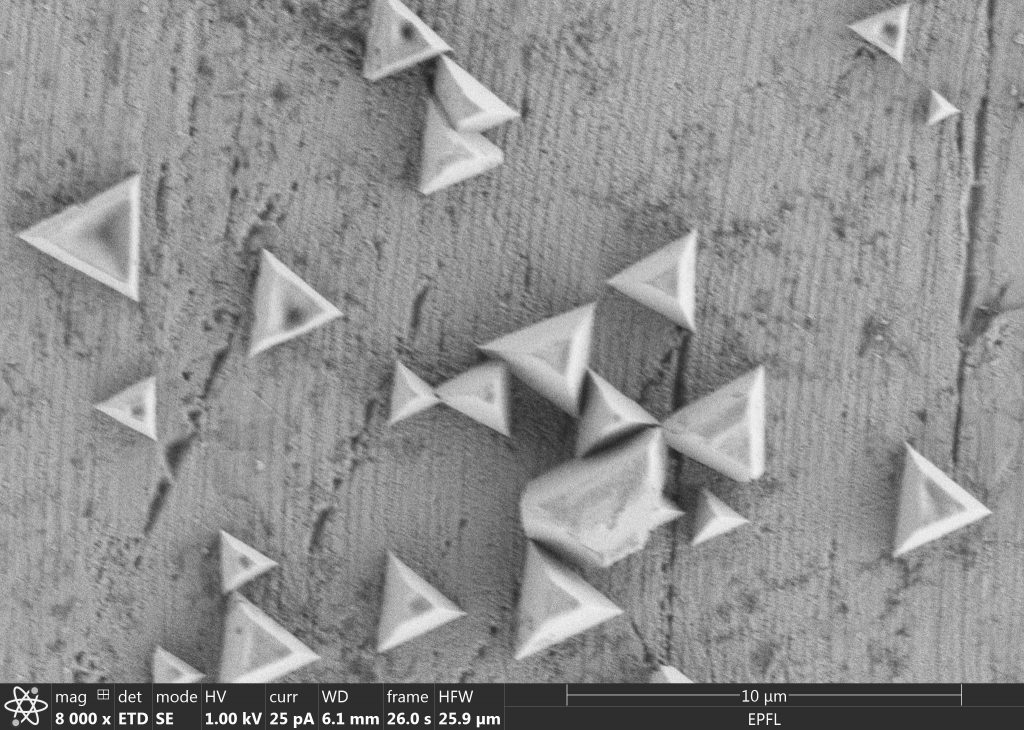
Carbon nitride crystals on copper substrate. Microscope: FEI Teneo SEM (Sion). 4/4
Polymeric microparticles processed via vortexing. Microscope: SEM Gemini.
Nothing better than a nano ice cream during a hot summer afternoon. Composed of randomly distributed Cu nanocrystals around a Cu rod. Microscope: TEM Tecnai G2 Spirit Twin (Sion).
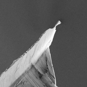
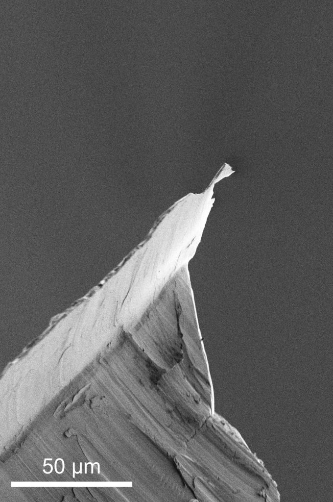
The probe apex (made by Pt/Ir wire) for the scanning tunneling microscope (Sion). Microscope: FEI Teneo SEM.
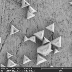
Carbon nitride crystals on copper substrate. Microscope: FEI Teneo SEM (Sion).
May/June 2022 competitors
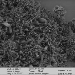
Here we see C-S-H + sulfates after 3 days, grown on the surface of calcite in its natural habitat. A predatory marine animal under the sea, C-S-H Anemone is the glue of cement on land. C-S-H anemones are classified in the phylum Cem-idaria, class Concretidum, and subclass Hydratea-cordoxum. Microscope: TEM Osiris 1/8
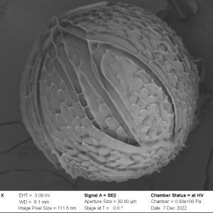
CaCO3 crystalizes (sometimes) into metastable vaterite crystals, that take unexpected shapes. Microscope: Gemini SEM 2/8
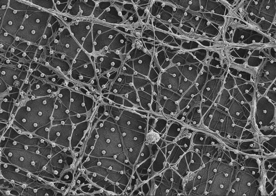
Primary hippocampal neurons growing on a single-crystal diamond chip. The surface is nanostructured with pillars having a diameter of 200nm and a height of 1 µm. The image is acquired after cells fixation and Au/Pd sputtering. Magnification x6000. Microscope: Gemini SEM 3/8
A spherical inclusion sitting on a chemically etched iron surface. Microscope: Gemini SEM 4/8
C-S-H with sulfate cradles and adorns our new calcite king and presents it to the animal kingdom. It’s the circle of life. And it moves us all. Microscope: TEM Osiris 5/8
Epoxy/Polycaprolactone blend microstructure after chloroform etching. Microscope: Gemini SEM 6/8
C-S-H with sulfates grown in the presence of calcite. Ribbons touch pinkies in the sky. Microscope: TEM Osiris 7/8
Primary hippocampal neurons growing on a single-crystal diamond chip. The surface is nanostructured with pillars having a diameter of 200nm and a height of 1 µm. The image is acquired after cells fixation and Au/Pd sputtering. Magnification x6000. Microscope: Gemini SEM 8/8
Here we see C-S-H + sulfates after 3 days, grown on the surface of calcite in its natural habitat. A predatory marine animal under the sea, C-S-H Anemone is the glue of cement on land. C-S-H anemones are classified in the phylum Cem-idaria, class Concretidum, and subclass Hydratea-cordoxum. Microscope: TEM Osiris
CaCO3 crystalizes (sometimes) into metastable vaterite crystals, that take unexpected shapes. Microscope: Gemini SEM

Primary hippocampal neurons growing on a single-crystal diamond chip. The surface is nanostructured with pillars having a diameter of 200nm and a height of 1 µm. The image is acquired after cells fixation and Au/Pd sputtering. Magnification x6000. Microscope: Gemini SEM
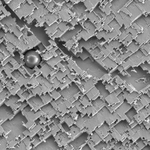
A spherical inclusion sitting on a chemically etched iron surface. Microscope: Gemini SEM
C-S-H with sulfate cradles and adorns our new calcite king and presents it to the animal kingdom. It’s the circle of life. And it moves us all. Microscope: TEM Osiris
Epoxy/Polycaprolactone blend microstructure after chloroform etching. Microscope: Gemini SEM
C-S-H with sulfates grown in the presence of calcite. Ribbons touch pinkies in the sky. Microscope: TEM Osiris
Primary hippocampal neurons growing on a single-crystal diamond chip. The surface is nanostructured with pillars having a diameter of 200nm and a height of 1 µm. The image is acquired after cells fixation and Au/Pd sputtering. Magnification x6000. Microscope: Gemini SEM
March/April 2022 competitors
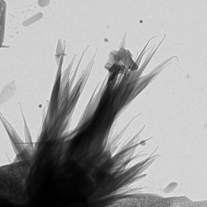
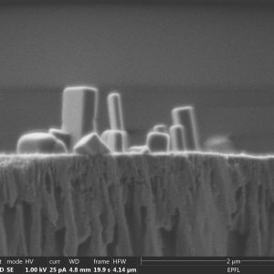
Germanium monsters arose at the bottom of a Silicon ocean from a Ion Beam Etching gone wrong. They are waiting with their gaping jaws for the next pray passing by. Microscope: SEM/FIB NVision. 1/10

Primary hippocampal neurons growing on a single-crystal diamond chip. The surface is nanostructured with pillars having a diameter of 200 nm and a height of 1µm. The image is taken on CIME’s Merlin microscope, after cells fixation and Au/Pd sputtering. Microscope: SEM Merlin. 2/10
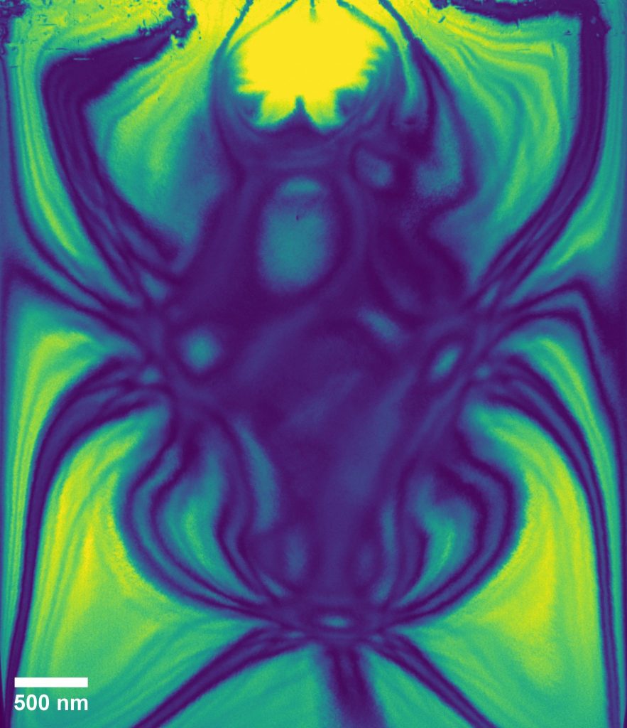
KNbO3 lamella FIB prepared. A spider appears on it as the effect of bend contours. Microscope: TEM Talos. 3/10
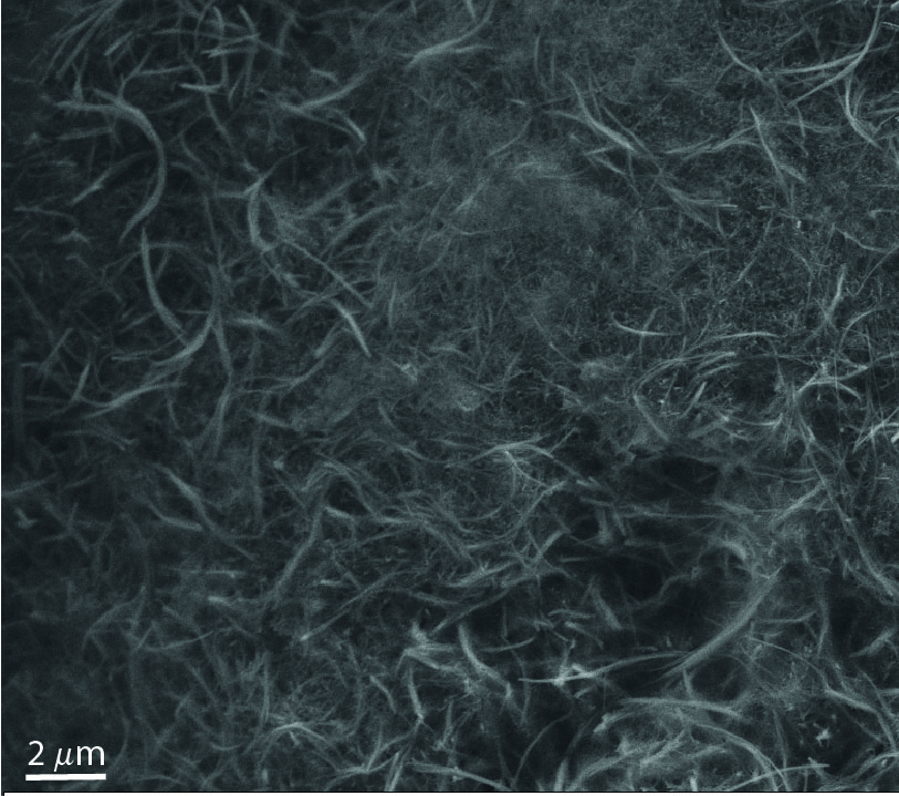
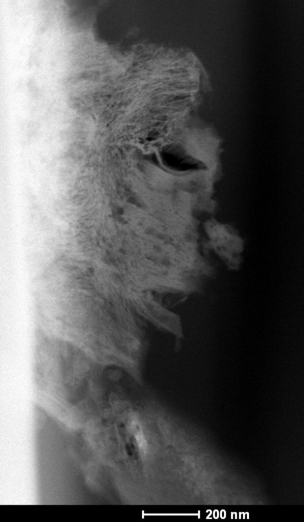
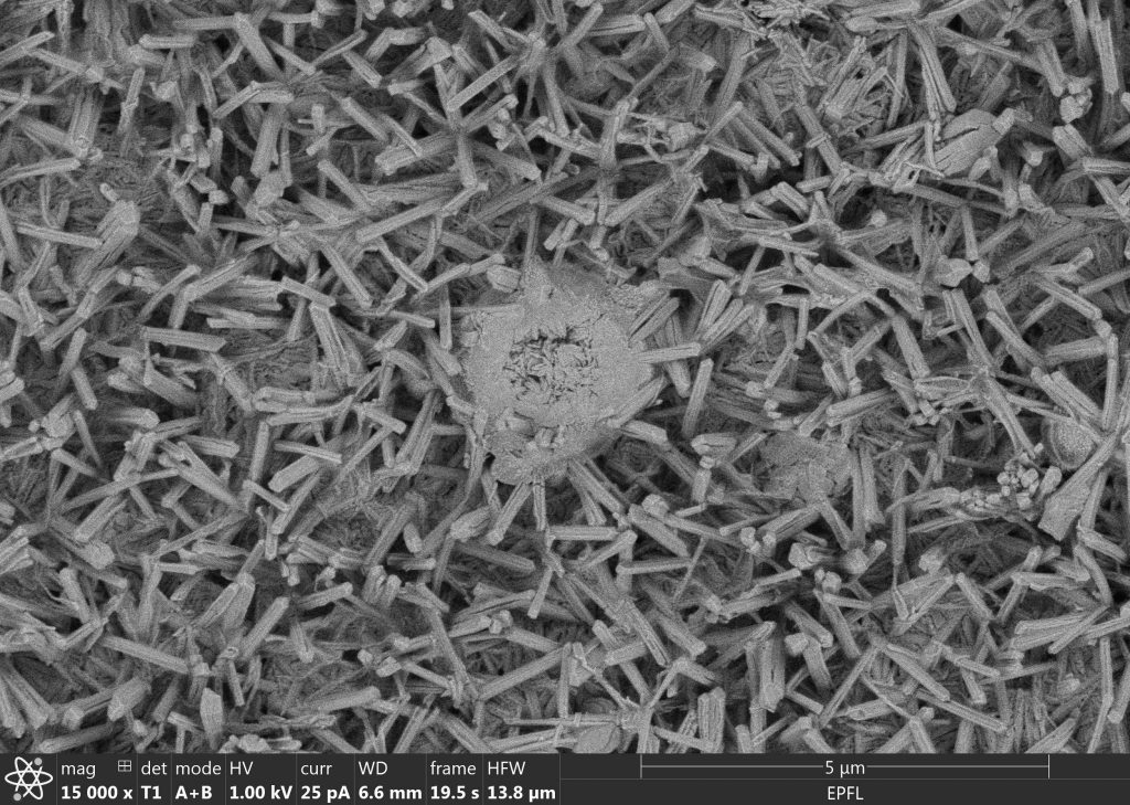
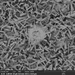
Blend of titanium dioxide nanofibers of different lengths. Microscope: SEM Merlin. 4/10
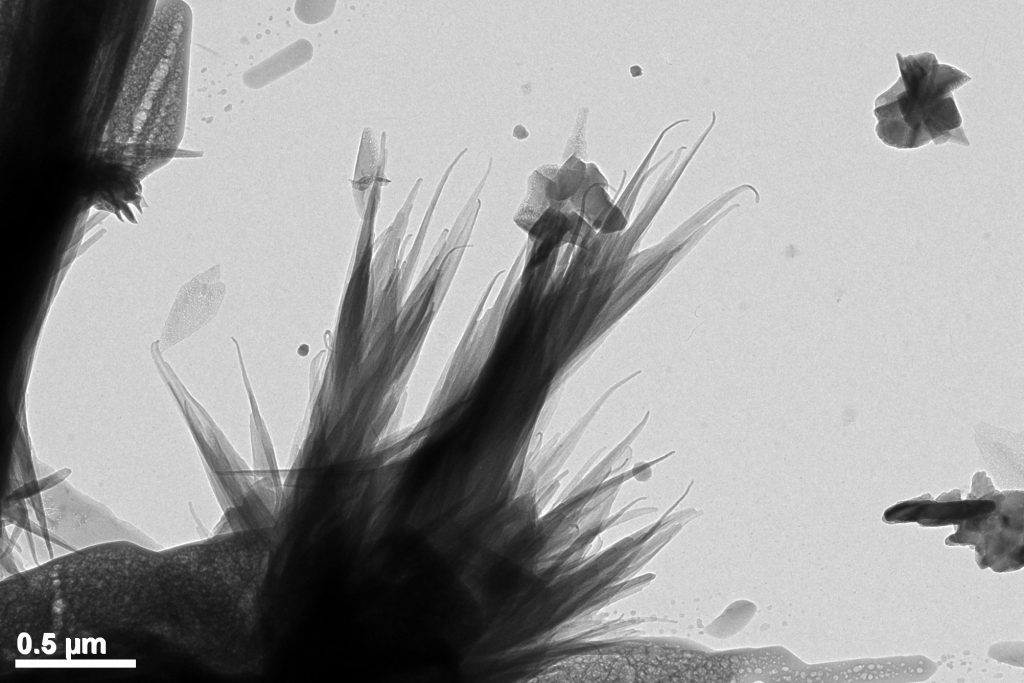
Natrolite crystals grown from grains of calcite in solution forming a tree adorned by portlandite and silica, ripe after only 3 days of reaction. Microscope: TEM Osiris. 5/10
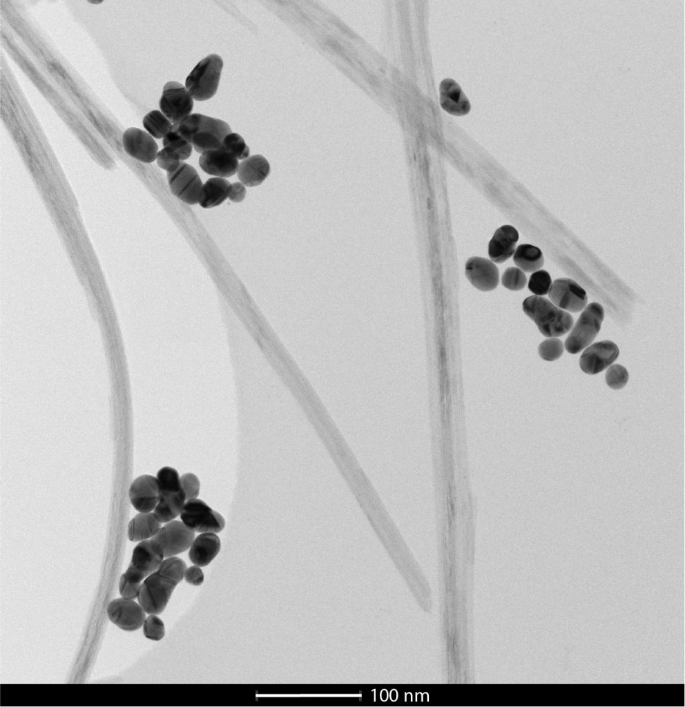
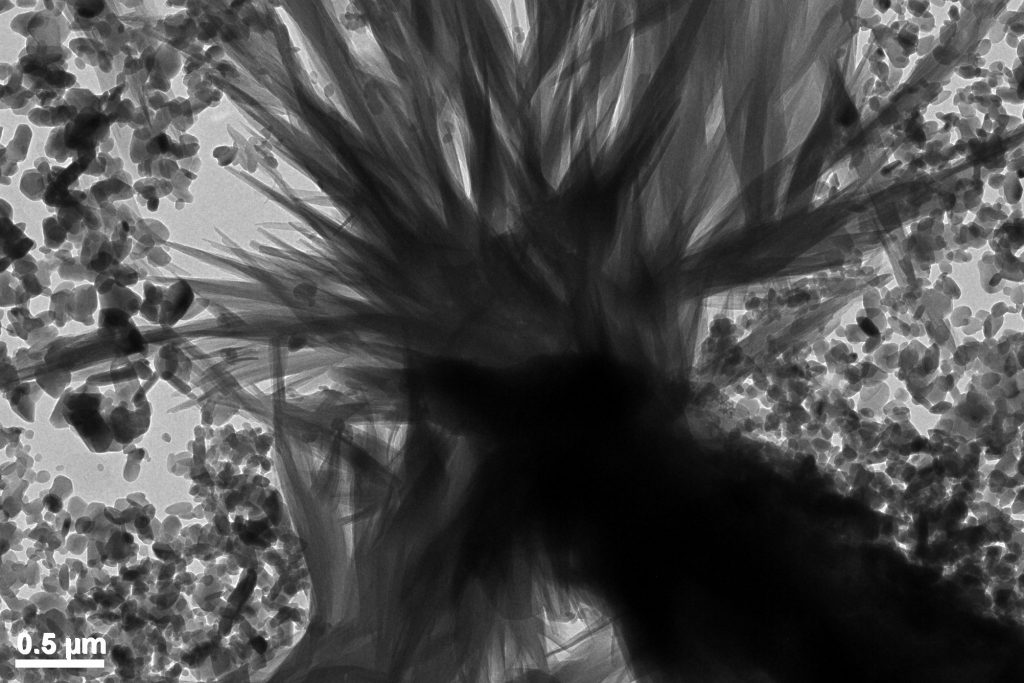
Gold nanoparticles on titanium dioxide nanofibers synthesized via sol-gel technique. Microscope: TEM Talos. 6/10
STEM-HAADF image of low carbon cement hydrated at 28 days. Microscope: TEM Osiris. 7/10
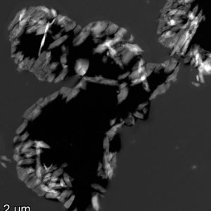
Aluminium metal-matrix composite with 20% in volume of SiC particles. Microscope: SEM Merlin. 8/10
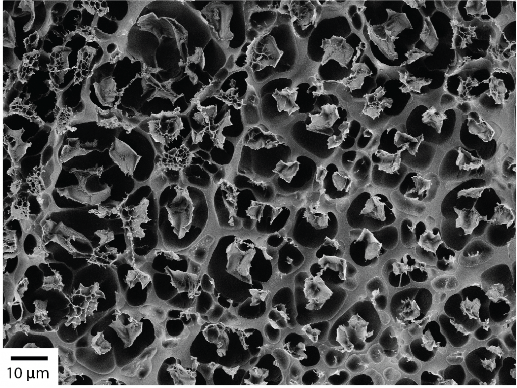
Double network granular hydrogel, fractured in two. Sputtered with 3nm Au/Pd, observed with SEM Merlin, SE2 detector. 9/10

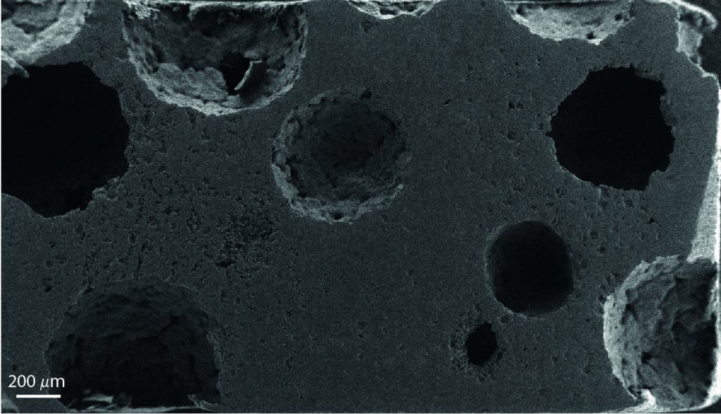
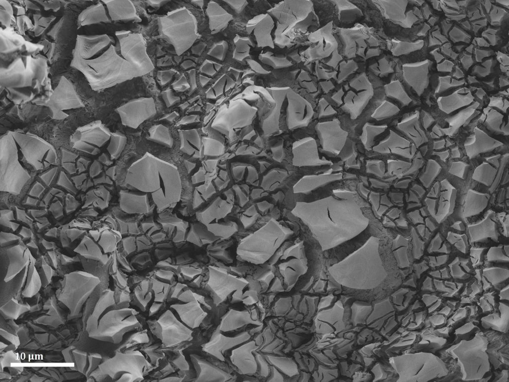
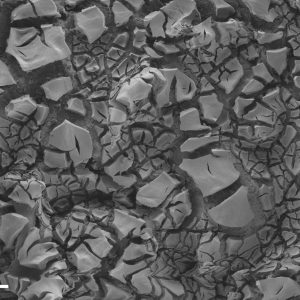
Spray deposition of catalyst particles on borosilicate-based support. Densification during the sintering step leads to vertical cracking of the coated particles. SEM image was taken on CIME’s Merlin microscope. 10/10
Germanium monsters arose at the bottom of a Silicon ocean from a Ion Beam Etching gone wrong. They are waiting with their gaping jaws for the next pray passing by. Microscope: SEM/FIB NVision.
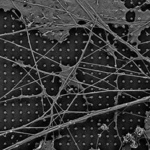
Primary hippocampal neurons growing on a single-crystal diamond chip. The surface is nanostructured with pillars having a diameter of 200 nm and a height of 1µm. The image is taken on CIME’s Merlin microscope, after cells fixation and Au/Pd sputtering. Microscope: SEM Merlin.
KNbO3 lamella FIB prepared. A spider appears on it as the effect of bend contours. Microscope: TEM Talos.
Blend of titanium dioxide nanofibers of different lengths. Microscope: SEM Merlin.
Natrolite crystals grown from grains of calcite in solution forming a tree adorned by portlandite and silica, ripe after only 3 days of reaction. Microscope: TEM Osiris.
Gold nanoparticles on titanium dioxide nanofibers synthesized via sol-gel technique. Microscope: TEM Talos.
STEM-HAADF image of low carbon cement hydrated at 28 days. Microscope: TEM Osiris.
Aluminium metal-matrix composite with 20% in volume of SiC particles. Microscope: SEM Merlin.
Double network granular hydrogel, fractured in two. Sputtered with 3nm Au/Pd, observed with SEM Merlin, SE2 detector.

Spray deposition of catalyst particles on borosilicate-based support. Densification during the sintering step leads to vertical cracking of the coated particles. SEM image was taken on CIME’s Merlin microscope.
January/February 2022 competitors
Spray deposition of catalyst particles on a borosilicate based support. Densification during the sintering step leads to vertical cracking of the coated particles. SEM image taken on CIME’s Merlin microscope. 1/6
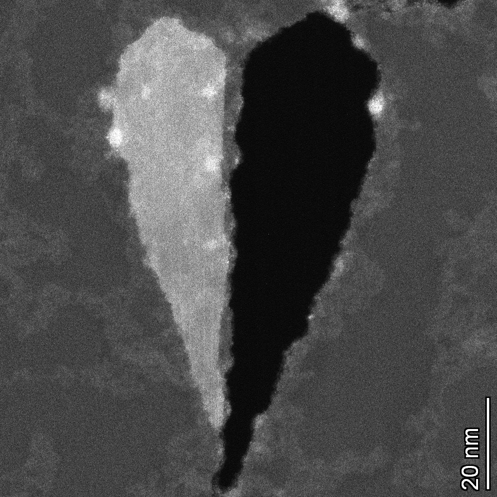
Tear in the MoS2 making heart shape. Microscope: TEM Titan Themis. 2/6
Transmission electron microscopy (TEM) image of zeolite powders. Microscope: TEM Osiris. 3/6
Calcium silicate hydrate (C-S-H) and ettringite coral in a cement sample after one day of hydration. Microscope: SEM Merlin. 4/6
We deposited the carbon nitride crystals onto an anodic aluminum oxide substrate. The SEM image of the cross-section was taken using SEM Teneo in Sion. 5/6
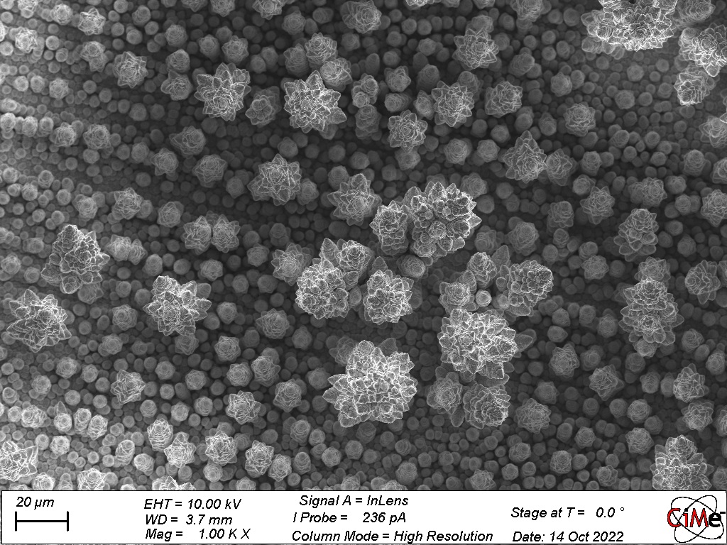
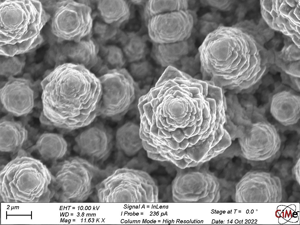
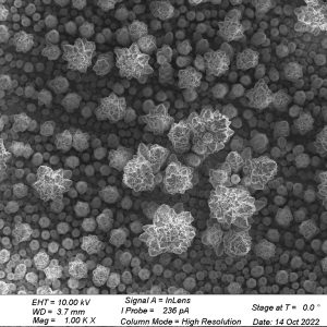
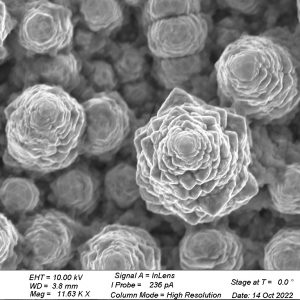
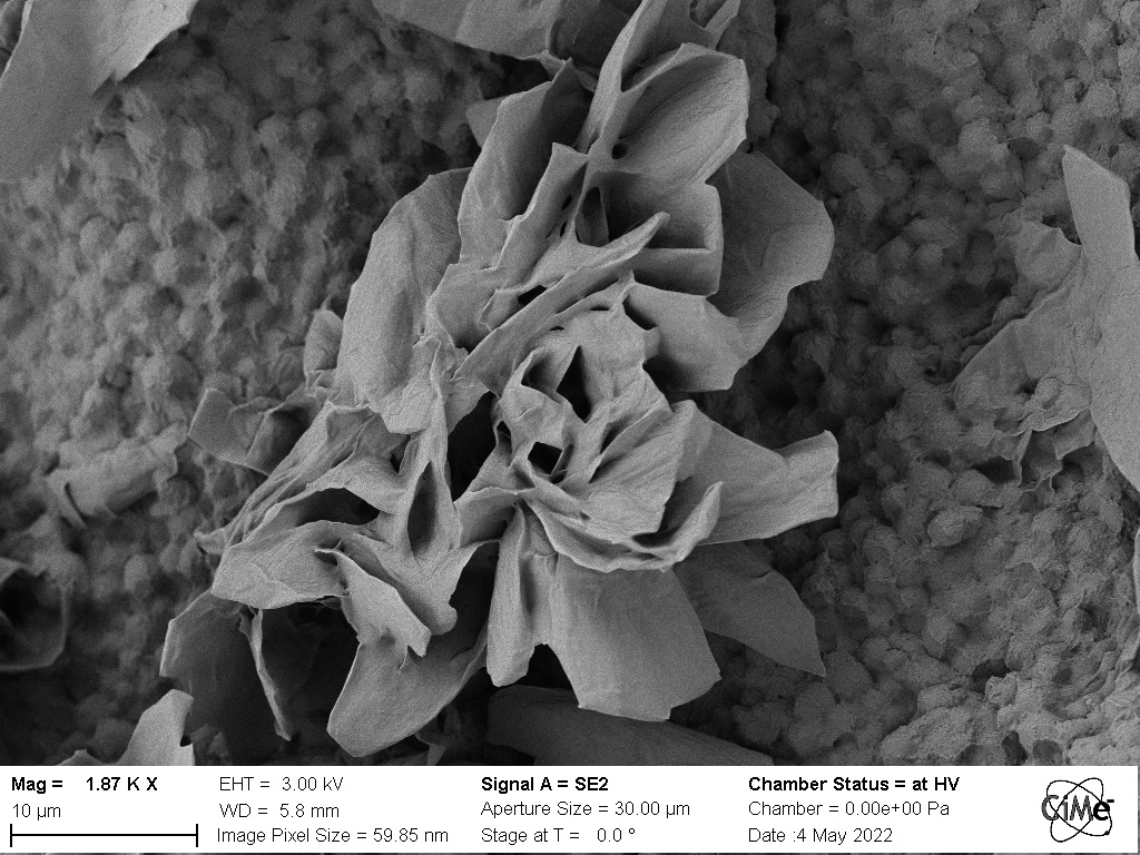
The SEM image of the as-synthesized crystalline carbon nitride blossom though the crystallinity was not good. The image was taken using SEM Teneo in Sion. 6/6
Spray deposition of catalyst particles on a borosilicate based support. Densification during the sintering step leads to vertical cracking of the coated particles. SEM image taken on CIME’s Merlin microscope.
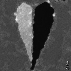
Tear in the MoS2 making heart shape. Microscope: TEM Titan Themis.
Transmission electron microscopy (TEM) image of zeolite powders. Microscope: TEM Osiris.
Calcium silicate hydrate (C-S-H) and ettringite coral in a cement sample after one day of hydration. Microscope: SEM Merlin.
We deposited the carbon nitride crystals onto an anodic aluminum oxide substrate. The SEM image of the cross-section was taken using SEM Teneo in Sion.
The SEM image of the as-synthesized crystalline carbon nitride blossom though the crystallinity was not good. The image was taken using SEM Teneo in Sion.